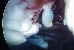
Earlier this Spring, I dislocated my patella, twice, about 2 weeks apart. This comes with some history, and was not the first time, but this time I decided it's time to do something about it.
And thus I have a whole video of these sort of interesting views, recorded last week during knee surgery. Although it's intellectually interesting, I'm still swollen and recovering, so it's actually slightly painful to watch the surgeon cleaning up, cauterizing and tightening the various parts of the inside of my knee. Maybe with some time, the pain will fade and it will just be interesting.
This image is from the before part, so, most of the loose blobs there were removed during the surgery.
As a data analyst, I'm intrigued by these sorts of images -- it takes a highly trained and experienced surgeon to really know what they are looking at. I could only guess, and it's my knee. If an untrained human can't really make heads or tails out of it, what sorts of knowledge would a computer need to "understand" it? Would there be a way to arthroscope a joint, with a single pass-through with a camera, then have a computer do an analysis of what's normal, what's not, and highlight some of those parts for a doctor in real-time, would that be of benefit? I suppose that as an academic, it's just and intriguing problem. But as an operational realist (and one with a bad knee), I wonder what those benefits would be and if such a technology might help me (or might have helped me).

 RSS Feed
